Search Index
355 results found
- Emotional chemistry on a molecular level | Scientia News
From bonds to emotions Facebook X (Twitter) WhatsApp LinkedIn Pinterest Copy link Emotional chemistry on a molecular level 26/06/25, 10:12 Last updated: Published: 16/01/24, 00:03 From bonds to emotions Emotions have a crucial role in how we perceive the world, behave, and interact with others. Our emotional states significantly influence how our lives are shaped, from the happiness of a long-awaited reunion to the grief of a heartbreaking farewell. But have you ever wondered what happens on a molecular level when we experience emotions? In this article, we will delve into the fascinating world of the chemistry behind emotions and explore how neurotransmitters, hormones, and brain regions collaborate to orchestrate the symphony of our feelings. Neurotransmitters , the chemical messengers in charge of transferring impulses between brain neurons, lie at the core of the chemistry of emotions. The "happiness hormone," serotonin , is known for its critical function in controlling mood, appetite, and sleep. Anxiety and sadness have been associated with low serotonin levels. Dopamine : this "reward neurotransmitter" is linked to reinforcement and pleasure. Dopamine is released when we like or receive a reward, which reinforces the behaviour and motivates us to seek out more of those kinds of experiences. Norepinephrine is a component of the body's fight-or-flight response that causes increased attention and arousal in reaction to stress or danger. Lastly, Gamma-Aminobutyric Acid (GABA) , an inhibitory neurotransmitter, counteracts the effects of excitatory neurotransmitters to maintain emotional stability by calming and soothing the brain. Our emotional experiences are significantly shaped by hormones as well. These chemical messengers affect the brain and other organs by entering the circulation after being released by numerous glands throughout the body. Cortisol , also referred to as the "stress hormone," is a key component of the body's fight-or-flight response and is released while under stress. Anxiety and a sense of being overpowered might result from elevated cortisol levels. The "love hormone" or "bonding hormone," oxytocin , is a chemical that is released during social interactions, particularly during times of closeness, trust, and bonding. The body's own natural mood lifters and painkillers are called endorphins . Exercise, laughing, and other enjoyable activities all produce endorphins, which contribute to a feeling of pleasure. Emotions are orchestrated within various brain regions , each with its own role in processing and interpreting emotional stimuli. Some key brain regions associated with emotions are: Amygdala : The "emotional hub" of the brain is commonly referred to as the amygdala. It analyses emotional inputs, particularly those connected to aggressiveness and fear, and participates in the development of emotional memories. Prefrontal cortex: This part of the brain controls rational higher-order thought, judgement, and emotional regulation. Even in highly emotional situations, it supports our ability to control our emotions and make logical decisions. Hippocampus : The hippocampus helps people remember emotional memories in particular. It is essential for remembering previous emotional experiences and creating emotional bonds. In conclusion, the chemistry of emotion is a gorgeously sophisticated dance of neurotransmitters, hormones, and different parts of the brain. It highlights the delicate balance that shapes our emotional experiences and influences our behaviour and well-being. Understanding this molecular magic can provide insight into our emotional reactions and open the door to novel treatment strategies for treating emotional disorders and mental health issues. Next time you feel overwhelmed with joy, anger, or any emotion in between, remember that there's a symphony of chemicals and brain activity behind the scenes, composing the unique melody of your emotional journey. Embrace your emotions, for they are an essential part of what makes us human. Written by Navnidhi Sharma Related articles: Exploring food at the molecular level / Psychology of embarrassment / Unmasking aggression / Chemistry of depression / Music and emotions Project Gallery
- Secondary bone cancer | Scientia News
Pathology and promising therapeutics Facebook X (Twitter) WhatsApp LinkedIn Pinterest Copy link Secondary bone cancer 11/07/25, 09:52 Last updated: Published: 13/12/23, 17:27 Pathology and promising therapeutics Introduction: what is secondary bone cancer? Secondary bone cancer occurs when cancer cells spread to the bones from a tumour that started somewhere else in the body. The site where the tumour first develops is called primary cancer. Cancer cells can break away from the primary cancer, travel through the bloodstream or lymphatic system, and establish secondary cancers, known as metastasis. Bones are among the most common sites to which cancer can spread. Most type of cancer has the potential to metastasise to the bones, with the most frequent occurrences seen in prostate, breast, lung, thyroid, kidney, and myeloma cancers. Throughout the literature, secondary cancer in the bones is referred to as bone secondaries or bone metastases. The most common areas of secondary bone cancer are the spine, ribs, pelvis, humerus (upper bone of the arm), femur (upper bone of the leg) and skull. There are two main types of bone cancer referred to as osteolytic and osteoblastic metastases. In osteolytic metastases, cancer cells break down the bone, leading to significant weakening. This type of metastasis is more common than osteoblastic metastases and often occurs when breast cancer spreads to the bone. In osteoblastic metastases, cancer cells invade the bone and stimulate excessive bone cell formation. This process results in the bone becoming very dense (sclerotic). Osteoblastic metastases frequently occur when prostate cancer spreads to the bone. Although new bone forms, it grows abnormally, which weakens the overall bone structure. Hormone therapy Like primary bone cancer, treatment for secondary bone cancer includes surgical excision, chemotherapy, and radiation therapy. Treatment for secondary bone cancer aims to control the cancer growth and symptoms. Treatment depends on several factors, including the type of primary cancer, previous treatment, the number of bones affected by cancer, whether cancer has spread to other body parts, overall health, and symptoms. Breast and prostate cancers rely on hormones for their growth. Reducing hormone levels in the body can be effective in managing the proliferation of secondary cancer. Hormone therapy, also known as endocrine therapy, uses synthetic hormones to inhibit the impact of the body’s innate hormones. Typical side effects include hot flashes, mood fluctuations, changes in weight, and sweating. Bisphosphonates Bone is a dynamic tissue with a continuous process of bone formation and resorption. Osteoclasts are cells responsible for breaking down bone tissue. In secondary bone cancer, cancer cells often produce substances that stimulate the activity of osteoclasts. This leads to elevated levels of calcium in the blood (hypercalcemia), resulting in feelings of nausea and excessive thirst. Treating secondary bone cancer involves strengthening bones, alleviating bone pain and managing hypercalcaemia). One option for bone-strengthening is bisphosphonates. Bisphosphonates can be administered orally or intravenously. They have been in clinical practice for over 50 years and are used to treat metabolic bone diseases, osteoporosis, osteolytic metastases, and hypercalcaemia. These compounds selectively target osteoclasts to inhibit their function. Bisphosphonates can be classified into two pharmacologic categories based on their mechanism of action. Nitrogen-containing bisphosphonates, the most potent class, function by suppressing the activity of farnesyl pyrophosphate synthase, a key factor in facilitating the binding of osteoclasts to bone. Consequently, this interference causes the detachment of osteoclasts from the bone surface, effectively impeding the process of bone resorption. Examples of these bisphosphonates include alendronate and zoledronate. Bisphosphonates without nitrogen in their chemical structure are metabolised intracellularly to form an analogue of adenosine triphosphate (ATP), known as 5'-triphosphate pyrophosphate (ApppI). ApppI is a non-functional molecule that disrupts cellular energy metabolism, leading to osteoclast cell death (apoptosis) and, consequently, reduced bone resorption. Examples of these bisphosphonates include etidronate and clodronate. Non-nitrogen-containing bisphosphonates can inhibit bone mineralisation and cause osteomalacia, a condition characterised by bones becoming soft and weak. Due to these considerations, they are not widely utilised. Denosumab Denosumab is another option for bone strengthening. It is administered as an injection under the skin (subcutaneously). Denosumab is a human monoclonal antibody that inhibits RANKL to prevent osteoclast-mediated bone resorption. Denosumab-mediated RANKL inhibition hinders osteoclast maturation, function, and survival in contrast to bisphosphonates, which bind to bone minerals and are absorbed by mature osteoclasts. In some studies, Denosumab demonstrated equal or superior efficacy compared to bisphosphonates in preventing skeletal-related events (SREs) associated with bone metastasis. Denosumab’s mechanism of action provides a targeted approach that may offer benefits for specific populations, such as patients with renal impairment. Bisphosphonates are excreted from the human body by the kidneys. A study by Robinson and colleagues demonstrated that bisphosphonate users had a 14% higher risk of chronic kidney disease (CKD) stage progression (including dialysis and transplant) than non-users. On the other hand, denosumab is independent of renal function and less likely to promote deteriorations in kidney function. Take-home message Secondary bone cancer, resulting from the spread of cancer cells to the bones, poses challenges across various cancers. Two main types, osteolytic and osteoblastic metastases, impact bone structure differently. Hormone therapy, bisphosphonates, and Denosumab have shown promising results and offer effective management of secondary bone cancers. Ultimately, the decision between treatments should be made in consultation with a healthcare professional who can evaluate the specific clinical situation and individual patient factors. The choice should be tailored to meet the patient’s needs and treatment goals. Written by Favour Felix-Ilemhenbhio Related article: Bone cancer Project Gallery
- Light: one of the biggest mysteries in physics | Scientia News
Simplifying light: photons, wave-particle duality and the Observer Effect Facebook X (Twitter) WhatsApp LinkedIn Pinterest Copy link Light: one of the biggest mysteries in physics Last updated: 20/10/25, 14:28 Published: 23/10/25, 07:00 Simplifying light: photons, wave-particle duality and the Observer Effect Light is one of those few topics where physicists have to say, ‘We don’t yet know why it is the way it is, we just know that it is that way.’ Let’s start simple. Question 1: What is light? When we think of light, we automatically think of visible light- what we can see with our eyes. But that is only 0.0035% of the total light, or electromagnetic, spectrum. The rest of the spectrum includes non-visible light, such as infrared radiation (what we feel as heat), x-rays (what the medical field’s X-ray machine uses to capture images of bones) or ultraviolet radiation (what causes sunburn). Every kind of light is made up of photons. They are tiny little pockets of energy that travel across space at 3 x 10 8 meters/second at different wavelengths and frequencies. Imagine someone tosses you a tennis ball, but instead of it travelling straight towards you, it oscillates up and down in a wave pattern as it travels. If you take a measurement from peak to peak, this distance is called a wavelength. The tennis ball can move up and down in the wave pattern at different speeds. This speed is called the frequency. Photons can travel at different wavelengths and different frequencies depending on where it originated. The unique wavelength and frequency pair of each photon determines what kind of light it is- where it falls on the electromagnetic spectrum. For example, photons with much shorter wavelengths and therefore much higher frequencies fall towards the right-hand side of the spectrum and are likely gamma-rays or x-rays. On the other hand, photons with much longer wavelengths and much lower frequencies are on the left-hand side, meaning the photons are probably radio waves or microwaves. So far, so good. All of this makes sense, and physicists are fairly confident in this information. So, what’s the problem? Question 2: Why is light so problematic? The trouble with light is its behaviour. Remember those little pockets of energy that move up and down in a wave pattern? Well, that’s not exactly what happens. Light has a property that physicists call ‘wave-particle duality’, which is a fancy term for meaning that sometimes light behaves like a particle (photons) and other times it behaves like a wave. When it behaves as a wave, we get the electromagnetic spectrum. As mentioned above, the wave can have different peak-to-peak lengths and travelling speeds that we read as different types of light across the spectrum. But when the photon behaves as a particle, we get this tiny pocket of energy rocketing across the cosmos. It is the fastest thing in the known universe. To understand the difference a little bit better, imagine you put the tennis ball in one of those pitching machines used for baseball players to practice their swing. It shoots the ball straight out of the front in a direct line and incredibly fast. This is light acting like a photon particle. Now, imagine you and a friend have a rope and each of you are holding on to either end. Your friend starts swinging their end up and down creating waves that travel down the rope towards you. The faster your friend swings their end, the faster the waves travel and the smaller the peak-to-peak distances (wavelengths) of the waves get, and vice versa if your friend slowly swings their end. This is light acting like a wave. The tricky bit is that physicists don’t know why the same pocket of energy can act like a photon particle in one instance, yet like a wave in another! The famous Double-Slit Experiment performed by Thomas Young in 1801 demonstrated this behaviour. Since then, the physics sub-field of quantum mechanics has developed and physicists now think that this behaviour is because of what they call the ‘Observer Effect’, which means that particles behave differently depending on whether or not they are observed. How does the particle know when it is being observed? Well, that is still a mystery to all. Written by Amber Elinsky Related articles: Laser Interferometric Gravitational-wave Observatory (LIGO) / Dark Energy Spectroscopic Instrument (DESI) REFERENCES Wavelength/Frequency Image ref: BYJU’s educational tech company Electromagnetic Image ref: Space.com Baclawski, Kenneth. (2018). The Observer Effect. 83-89. 10.1109/COGSIMA.2018.8423983. Project Gallery
- Depression in Children | Scientia News
Getting treatment can prevent things from getting worse Facebook X (Twitter) WhatsApp LinkedIn Pinterest Copy link Depression in Children 10/07/25, 10:17 Last updated: Published: 17/06/23, 12:46 Getting treatment can prevent things from getting worse It's normal for kids to feel sad, act grouchy, or be in a bad mood at times. But when a sad or bad mood lasts for weeks or longer, and when there are other changes in a child's behavior, it might be depression. Therapy can help children who are going through sadness or depression. And there are things parents can do, too. Getting the right care can prevent things from getting worse and help a child feel better. Symptoms of depression Sad or bad mood. A child may seem sad, lonely, unhappy, or grouchy. It can last weeks or months. A child may cry more easily. They may have more tantrums than before. Being self-critical. Kids going through depression may complain a lot. They may say self-critical things like, "I can't do anything right." "I don't have any friends." "I can't do this." "It's too hard for me." Lack of energy and effort. Depression can drain a child's energy. They might put less effort into school than before. Even doing little tasks can feel like too much effort. Kids may seem tired, give up easily, or not try. Not enjoying things. Kids don't have as much fun with friends or enjoy playing like before. They may not feel like doing things they used to enjoy. Sleep and eating changes. Kids may not sleep well or seem tired even if they get enough sleep. Some may not feel like eating. Others may overeat. Aches and pains. Some children may have stomach aches or other pains. Some miss school days because of not feeling well, even though they aren't sick. Causes of depression Some common reasons include: - life events like someone dying - moving schools or other big changes - physical health problems - experiencing physical, sexual or psychological abuse or neglect - witnessing violence or a traumatic event - if you have an unstable family environment Intervention Three of the more common methods used in depression treatment include: - cognitive behavioral therapy - interpersonal therapy - psychodynamic therapy Written by Chhaya Dhedi Related articles: Childhood stunting in developing nations / What does depression do to your brain? / Brain of a bully / Anxiety / Postpartum depression in adolescent mothers Project Gallery
- The environmental impact of EVs | Scientia News
A chemical perspective Facebook X (Twitter) WhatsApp LinkedIn Pinterest Copy link The environmental impact of EVs 16/01/25, 11:21 Last updated: Published: 07/08/23, 15:58 A chemical perspective Electric vehicles (EVs) are gaining momentum worldwide as a greener alternative to conventional internal combustion engine vehicles (ICEVs). The environmental benefits of EVs extend beyond their efficient use of electricity. In this article, we explore the chemical aspects of EVs and their environmental impact, shedding light on their potential to mitigate climate change and reduce pollution. Greenhouse Gas Emissions Reduction: EVs play a crucial role in addressing climate change by significantly reducing greenhouse gas (GHG) emissions. Unlike ICEVs that rely on fossil fuels, EVs generate zero tailpipe emissions. By utilising electricity as their energy source, EVs minimise the release of carbon dioxide (CO2) and other GHGs responsible for global warming. However, it's essential to consider the environmental implications of electricity generation, emphasising the need for renewable energy sources to maximise the positive impact of EVs. Battery Chemistry and Resource Management: The heart of an EV lies in its rechargeable battery, typically composed of lithium-ion technology. The production and disposal of these batteries present both opportunities and challenges. Raw materials, such as lithium, cobalt, and nickel, are essential components of EV batteries. Responsible mining practices and efficient recycling techniques are vital to minimising the environmental impact of resource extraction and ensuring proper disposal or repurposing of used batteries. Electrochemical Reactions and Energy Storage: Electric vehicles rely on electrochemical reactions within their batteries to store and release energy. These reactions involve the flow of ions, typically lithium ions, between the positive and negative electrodes. Understanding the chemistry behind these processes enables the development of more efficient and durable battery systems. Continued research and innovation in battery chemistry hold the potential to enhance energy storage capabilities, extend EV range, and improve overall performance. Air Quality and Emission Reduction: EVs contribute to improved air quality due to their zero tailpipe emissions. By eliminating the release of pollutants such as nitrogen oxides (NOx), particulate matter (PM), and volatile organic compounds (VOCs), EVs reduce smog formation and respiratory health risks. This is particularly significant in urban areas, where high concentrations of vehicular emissions contribute to air pollution. The adoption of EVs can help combat these issues and create cleaner and healthier environments. Battery Recycling and the Circular Economy: Given the increasing demand for EVs, battery recycling plays a vital role in ensuring a sustainable future. Recycling allows for the recovery of valuable materials and reduces the need for resource extraction. Effective recycling processes can mitigate the environmental impact of battery production, minimise waste generation, and promote a circular economy approach, where materials are reused and recycled to their fullest extent. Future Prospects and Chemical Innovations : Advancements in battery technology and chemical engineering are key to unlocking the full potential of EVs. Research efforts are focused on developing alternative battery chemistries, such as solid-state batteries, which offer improved energy density, safety, and recyclability. Additionally, exploring sustainable materials and manufacturing processes for batteries can further reduce the environmental footprint of EVs. In conclusion, electric vehicles represent a promising solution to combat climate change, reduce pollution, and promote sustainable transportation. From the chemistry behind battery systems to their impact on air quality and resource management, EVs offer a greener alternative to traditional vehicles. Continued research, innovation, and collaboration between the automotive industry, chemical scientists, and policymakers are essential for realising the full potential of EVs and creating a cleaner, more sustainable future. Written by Navnidhi Sharma Related articles: Hydrogen cars / The brain-climate connection / Plastics and their environmental impact Project Gallery
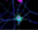
- Synaptic plasticity | Scientia News
Synaptic plasticity is the process of connections within the brain changing to adapt to new information over time. It is of increasing significance in neuroscience, especially in the field of memory. Early research into synaptic plasticity was conducted by many of those Go Back Facebook X (Twitter) WhatsApp LinkedIn Pinterest Copy link Our understanding of how the brain forms connections between things we’ve learnt, and how London taxi drivers fit in Last updated: 13/11/25 Published: 05/01/23 Synaptic plasticity is the process of connections within the brain changing to adapt to new information over time. It is of increasing significance in neuroscience, especially in the field of memory. Early research into synaptic plasticity was conducted by many of those now considered the pioneers of neuroscience. For instance, Terje Lomo (1966) experimented on rabbit hippocampus with repeated, high-frequency stimulation, identifying long term potentiation, the persistent strengthening of synapses leading to enduring increases in signal transmission between neurons. Prior to work by Lomo, Ramon y Cajal (1911) proposed the idea that the strength of synaptic connections had to change to alter existing memories. One key question which is pertinent for both humans and other animals alike is how to keep track of our surroundings - how do our memories encode and store information on the places we visit so we can remember the directions for next time? Seminal work by Maguire et al., (2001) assessed whether physical changes “could be detected in the healthy brain” of London taxi drivers, given the repertoire of spatial experience required to navigate London without aid. Sixteen taxi drivers were studied with fifty controls. Using (structural) magnetic resonance imaging (MRI), it was found that taxi drivers’ posterior hippocampi were larger than that of control subjects, and the more experience the drivers had, the greater the size of their right posterior hippocampi. Such changes in tissue volume take place gradually over time, because of task-related training. Recent work by Spiers et al., (2022) looked at the difference in spatial navigational ability between city-dwellers and those living in rural areas. A subset of ~400,000 participants from 38 countries played a video game to test their skill in spatial navigation, with city-dwellers performing worse than those who grew up outside cities. More specifically, individuals were better at navigating in environments that were topologically like where they grew up. Hence, one interpretation of these results is that the place where a person grows up impacts their ability to accurately navigate new, unfamiliar environments since this is based on the synaptic connections made between existing information in the brain. In conclusion, synaptic plasticity is the change in connections in the brain over time; interest and research in this field, especially spatial navigation, are increasing significantly. Written by Manisha Halkhoree Full article published in Brain Insights- BNA Bulletin (Issue no. 96, Autumn 2022) Related articles: The wonders of the human brain / The brain-climate connection / Why brain injuries affect adults and children differently REFERENCES Nicoll, R. A Brief History of Long-Term Potentiation. Neuron. 2017 Jan 18; 93(2): 281-290. Available from: https://www.sciencedirect.com/science/article/pii/S0896627316309576 Maguire E, Gadian D, Johnsrude I, et. al. Navigation-related structural change in the hippocampi of taxi drivers. Proc Natl Sci U S A. 2000 Apr 11; 97(8): 4398–4403. Available from: https://www.ncbi.nlm.nih.gov/pmc/articles/PMC18253/ Coutrot A, Manley E, Goodroe S., et. al. Entropy of city street networks linked to future spatial navigation ability. Nature. 2022 March 30; Nature 604: 104-110. Available from: https://www.nature.com/articles/s41586-022-04486-7
- 'The Emperor of All Maladies' by Siddhartha Mukherjee | Scientia News
Book review Facebook X (Twitter) WhatsApp LinkedIn Pinterest Copy link 'The Emperor of All Maladies' by Siddhartha Mukherjee 08/01/26, 18:58 Last updated: Published: 28/11/24, 14:55 Book review Stretching nearly 4,000 years of history, Pulitzer Prize winner Siddhartha Mukherjee sets on a journey to document the biography of cancer in The Emperor of All Maladies. Drawing from a vast array of books, studies, interviews, and case studies, Mukherjee crafts a narrative that is as comprehensive as it is compelling. Driven by curiosity and a desire to understand the origins of cancer, Mukherjee sets the tone by reflecting on his experiences as an oncology trainee, drawing insightful parallels to contemporary perspectives on the fight against this relentless disease. Mukherjee also pays homage to Ancient Egyptian and Greek physicians for their early observations on cancer, from the work on Imhotep to Claudius Galen. He then introduces Sidney Farber, whose monumental contributions to modern chemotherapy are brought to life through Mukherjee's exceptional storytelling—tracing Farber's journey from his initial observations to his unprecedented success in treating children with leukaemia. As you progress through each chapter of this six-part book, your appreciation deepens for how far cancer treatments have advanced - and how much further they can go. Mukherjee’s unparalleled skill as a science communicator shines through, seamlessly weaving together groundbreaking scientific discoveries with the historical contexts in which they emerged contributing to an immersive reading experience. Siddhartha Mukherjee, The Emperor of All Maladies : In 2005, a man diagnosed with multiple myeloma asked me if he would be alive to watch his daughter graduate from high school in a few months. In 2009, bound to a wheelchair, he watched his daughter graduate from college. The wheelchair had nothing to do with his cancer. The man had fallen down while coaching his youngest son's baseball team. Mukherjee also makes an effort to highlight the critical role of raising awareness in shaping public health outcomes. ‘Jimmy’ was a cancer patient that represented children with cancer, his real name was Einar Gustafson, but his individual story was able to galvanise large-scale support. As the face of the ‘Jimmy Fund’, he was able to assist in raising $231,485.51 for the Dana-Farber Institute subsequently becoming the official charity for the Boston Red Sox. Mukherjee underscores how storytelling can serve as a catalyst for change, not just in raising money, but also in enacting larger societal and governmental shifts. In 1971, President Richard Nixon signed the National Cancer Act, the first of its kind where federal funding went directly into advancing cancer research. What struck me most was how Mukherjee connects this historical event to the broader need for advocacy, as science doesn’t just happen in the lab. It is a collective effort, driven by awareness, to push funding and influence policy. The ability to link individual stories to broader missions, as Mukherjee illustrates, continues to be one of the most effective strategies in keeping cancer research in the public eye. Mukherjee delves into the pivotal role of genetics in cancer research, tracing its evolution from the discovery of DNA's structure by Francis Crick, James Watson, and Rosalind Franklin to Robert Weinberg's ground-breaking work on how proto-oncogenes and tumour suppressors drive cancer progression. These discoveries ushered in a new era in cancer drug development. Mukherjee also emphasises the importance of collaboration and the rise of the internet, which gave birth to The Cancer Genome Atlas, a landmark program, that unites various research disciplines to diagnose, treat, and prevent cancer. In concluding the book, Mukherjee looks ahead to the future of cancer treatment, seamlessly connecting this discussion to his second book, The Gene . This book takes readers on a remarkable journey through the history of cancer, from the earliest recorded cases to groundbreaking discoveries in genetics. It weaves together compelling personal stories as well as pivotal moments in governmental policy. The storytelling is rich and immersive, drawing you in with its detail and depth. By the time you finish, you'll find yourself returning to its pages, eager to revisit the knowledge and insights it offers. Written by Saharla Wasarme Related book reviews: Intern Blues / The Molecule Project Gallery
- Digital innovation in rural farming | Scientia News
Transforming agriculture with computer science Facebook X (Twitter) WhatsApp LinkedIn Pinterest Copy link Digital innovation in rural farming 09/07/25, 14:04 Last updated: Published: 21/07/23, 09:58 Transforming agriculture with computer science With their rich agricultural heritage and significant contribution to the national economy, rural farming communities have always been at the forefront of agricultural innovation. Today, as the world undergoes rapid digital transformation, the integration of computer science has emerged as a game-changer in the agricultural sector. By harnessing the power of emerging technologies and data-driven approaches, farmers can enhance productivity, optimize resource allocation, and foster sustainable farming practices. This article delves into the role of computer science in revolutionising agriculture and farming practices in rural areas. From precision agriculture and data analytics to the utilisation of IoT, drones, and decision support tools, we explore how technology-driven solutions are shaping a new era of agriculture, promising increased efficiency, reduced environmental impact, and improved livelihoods for farmers. A recent report revealed that farmers in various regions, specifically rural and eastern regions such as Punjab, India have faced significant challenges, including crop failures, leading to distress and financial difficulties. It is important to address these issues and prevent the associated consequences. Digitalisation within the farming industry can play a vital role in mitigating these challenges and fostering resilience. So how exactly can rural farming benefit from digitalisation? Precision agriculture and data analytics: the implementation of precision agriculture techniques, supported by data analytics, can enable farmers to optimise resource utilisation, improve crop management, and mitigate agricultural risks. By analysing data related to weather patterns, soil conditions, and crop health, farmers can make informed decisions, enhance productivity, and reduce the incidence of crop failures. Market intelligence and price forecasting: computer science tools can facilitate better market intelligence and price forecasting, empowering farmers to make informed decisions about crop selection, timing of harvest, and market strategies. Access to real-time market data, coupled with predictive analytics, can help farmers negotiate fair prices and reduce financial vulnerability caused by market instability. Remote sensing and drone technology: utilising remote sensing and drone technology can enable efficient crop monitoring, early detection of diseases, and targeted interventions. High-resolution imagery and computer vision algorithms can identify crop stress, nutrient deficiencies, or pest outbreaks, allowing farmers to take timely action, reduce crop losses, and enhance yield. Decision support systems: the introduction of decision support systems can provide customised recommendations to farmers, incorporating data from multiple sources such as weather forecasts, market trends, and agronomic best practices. These systems can assist farmers in making well-informed decisions regarding crop selection, input usage, and resource allocation, ultimately improving their profitability, and reducing financial distress. The integration of computer science offers promising avenues for addressing the complex challenges faced by farmers in rural areas. By harnessing the power of data analytics, IoT, drones, and decision support tools, farmers can benefit from enhanced agricultural practices, improved market access, and financial stability. However, it is crucial to ensure the accessibility and affordability of these technologies, coupled with comprehensive support systems and policy reforms, to truly empower farmers and create sustainable change. Written by Jaspreet Mann Related articles: Revolutionising sustainable agriculture through AI / Plant diseases and nanoparticles Project Gallery
- Nanogels: the future of smart drug delivery | Scientia News
Nanogels are tiny, water swollen polymer networks and encapsulate therapeutic agents Facebook X (Twitter) WhatsApp LinkedIn Pinterest Copy link Nanogels: the future of smart drug delivery Last updated: 17/07/25, 10:54 Published: 17/07/25, 07:00 Nanogels are tiny, water swollen polymer networks and encapsulate therapeutic agents Nanomedicine is a rapidly advancing field, with nanogels emerging as promising innovations for drug delivery applications. Nanogels are soft nanoscale hydrogels that are transforming how we deliver drugs and treat diseases. Whilst hydrogels themselves have long been used in biomedical applications such as tissue engineering and wound healing, their relatively larger sizes (above 100 micrometres) limits their ability to interact with cells and cross biological barriers. Nanogels, however, are thousands of times smaller, and offer unique advantages as a result. What are nanogels? Nanogels are tiny, water swollen polymer networks and are made up of crosslinked polymer chains to form a 3D matrix. Nanogels can encapsulate therapeutic agents inside their porous core shell structure. This swelling allowing nanogels to carry payloads, such as drugs, proteins, nucleic acids and these cargo materials are protected from degradation in the body whilst enabling controlled and targeted delivery. Due to their small sizes, nanogels can penetrate tissues and even enter cells, which overcomes the limitations faced with hydrogels. The surface of nanogels can also be engineered for specificity, to allow for precise targeting of drugs to receptors on diseased cells or inflamed tissues. Advantages over other nanocarriers Compared to liposomes and polymeric micelles, nanogels have a larger inner surface, which means they can carry more payload. The higher loading capacity improves the therapeutic efficiency whilst reducing the risks of side effects cause by off-target drug release. Nanogels also undergo the enhanced permeability and retention (EPR) effect - a phenomenon where the nanoparticles naturally accumulate in tumour or inflamed tissues due to leaky blood vessel, and as a result this improves drug delivery to targeted disease sites. Stimuli responsive ‘smart’ nanogels A key feature of nanogels is their stimuli responsiveness, or ability to act as ‘smart’ materials. The nanogels can be designed to respond to environmental triggers such as changes in pH, temperature, light, redox conditions, pressure and more. This responsiveness enables controlled release of drugs exactly when and where they are needed12. For example, thermoresponsive nanogels can change their structure at body temperature or when exposed to localised heating, making them ideal for applications like wound healing and cancer therapy. This controlled release prevents premature drug leakage, reduces systemic toxicity and overall improves the precision of the treatment. The future of nanogels in medicine Nanogels have huge potential as customisable drug delivery systems to target specific disease systems. They are biocompatible, stable, and have high drug loading capacities and are stimuli responsive; these properties combined make them a powerful tool in applications such as targeted drug delivery and gene therapy. As nanomedicine research progresses, nanogels are set to revolutionise healthcare with smarter, safer and more targeted therapies. Written by Saanchi Agarwal Related articles: Nanomedicine / Nanoparticles and diabetes treatment / Nanoparticles and health / Nanocarriers / Silicon hydrogel REFERENCES L. Blagojevic and N. Kamaly, Nanogels: A chemically versatile drug delivery platform, Nano Today, 2025, 61, 102645. F. Carton, M. Rizzi, E. Canciani, G. Sieve, D. Di Francesco, S. Casarella, L. Di Nunno and F. Boccafoschi, Use of Hydrogels in Regenerative Medicine: Focus on Mechanical Properties, Int. J. Mol. Sci. , 2024, 25 , 11426. N. Rabiee, S. Hajebi, M. Bagherzadeh, S. Ahmadi, M. Rabiee, H. Roghani-Mamaqani, M. Tahriri, L. Tayebi and M. R. Hamblin, Stimulus-Responsive Polymeric Nanogels as Smart Drug Delivery Systems, Acta Biomater. , 2019, 92 , 1–18. N. Rabiee, S. Hajebi, M. Bagherzadeh, S. Ahmadi, M. Rabiee, H. Roghani-Mamaqani, M. Tahriri, L. Tayebi and M. R. Hamblin, Stimulus-Responsive Polymeric Nanogels as Smart Drug Delivery Systems, Acta Biomater. , 2019, 92 , 1–18. A. Vashist, G. P. Alvarez, V. A. Camargo, A. D. Raymond, A. Y. Arias, N. Kolishetti, A. Vashist, P. Manickam, S. Aggarwal and M. Nair, Recent advances in nanogels for drug delivery and biomedical applications, Biomater. Sci. , 2024, 12 , 6006–6018. K. S. Soni, S. S. Desale and T. K. Bronich, Nanogels: an overview of properties, biomedical applications and obstacles to clinical translation, J. Control. Release Off. J. Control. Release Soc. , 2016, 240 , 109–126. A. Bordat, T. Boissenot, J. Nicolas and N. Tsapis, Thermoresponsive polymer nanocarriers for biomedical applications, Adv. Drug Deliv. Rev. , 2019, 138 , 167–192. K. S. Soni, S. S. Desale and T. K. Bronich, Nanogels: an overview of properties, biomedical applications and obstacles to clinical translation, J. Control. Release Off. J. Control. Release Soc. , 2016, 240 , 109–126. T. Alejo, L. Uson, G. Landa, M. Prieto, C. Yus Argón, S. Garcia-Salinas, R. de Miguel, A. Rodríguez-Largo, S. Irusta, V. Sebastian, G. Mendoza and M. Arruebo, Nanogels with High Loading of Anesthetic Nanocrystals for Extended Duration of Sciatic Nerve Block, ACS Appl. Mater. Interfaces , 2021, 13 , 17220–17235. S. V. Vinogradov, Nanogels in The Race for Drug Delivery, Nanomed. , 2010, 5 , 165–168. Project Gallery
- Same-sex attraction in non-human animals | Scientia News
Behaviours in birds, mammals, and invertebrates Facebook X (Twitter) WhatsApp LinkedIn Pinterest Copy link Same-sex attraction in non-human animals Last updated: 17/06/25, 11:20 Published: 11/09/25, 07:00 Behaviours in birds, mammals, and invertebrates Biased science communication can have detrimental consequences. For example, facts about animal reproduction have been twisted to justify discrimination against the LGBTQIA+ community. Some people call homosexuality a “Darwinian paradox”, because it does not fit their preconceived belief that an animal’s job is to stay alive and make babies. This belief ignores how some animals, like humans, have complex social structures and do things just for fun. Same-sex sexual behaviours (SSSB) have been observed in 1500 animal species, none of whom do it to make babies. This article describes some of these behaviours in birds, mammals, and invertebrates. Same-sex sexual behaviour (SSSB) in birds The first recorded example of SSSB in non-human animals comes from Aristotle about 2300 years ago. He wrote about male pigeons, partridges, and quails mating with other male conspecifics. Since then, same-sex relationships have been recorded in other bird species. Greylag geese form “gander pairs” of two males, whose behaviours resemble pairs of opposite-sex mates. In Oahu, Hawaii, female-female Laysan albatross pairs looked after 31% of nests between 2004 and 2007. These pairs, one of which is pictured in Figure 1 , were equally good at raising chicks as male-female pairs. SSSB was also observed in unbonded king penguins, meaning penguins which had not committed to a mate for that breeding season. Using DNA to assess individual sex, 26.4% of courtship displays between unbonded king penguin couples were same-sex. There was also one male-male and one female-female pair of bonded king penguins, but both couples broke up and re-bonded with opposite-sex mates in the same season. The most famous same-sex bird couple is Roy and Silo from Central Park Zoo. They were a pair of chinstrap penguins who raised a chick named Tango when given a fertile egg. This family was the subject of a children’s book ( Figure 2 ) and an American culture war. Thus, many bird species pair with individuals of the same sex in captivity and more importantly, in the wild. SSSB in mammals Humans are not the only mammals to mate with individuals of the same sex. Male bats from the Myotis genus have been observed getting intimate with each other, and Mytois lucifugus releases sperm during this activity. In another bat species called the Bonin flying fox, males groomed each other in a way scientists perceived as sexual. Japanese macaques have monogamous female-female pairs called consortships, in which females carry out the same mating behaviours seen with male-female pairs. SSSB in insects In addition to birds and mammals, some insects conduct sexual activities to others of the same sex. In a 2012 study, 16% of male field crickets did courtship displays to and/or tried to mate with another male. The authors conducted experiments to rule out some leading Darwinian causes of SSSB, such as establishing dominance relationships (similar to an ‘alpha male’) or defusing hostile encounters. SSSB is well studied in flour beetles, where the males mount other males and release capsules of sperm like they would to females. In these beetles, the sexes are sexually dimorphic - distinguishable by appearance, smell, and/or sound - so a male beetle is intentionally choosing to mate with another male. When 59 male damselflies were offered a male and female in the same cage, 10 approached and began mating with the male. More damselflies chose the male over the female after spending a few days in a male-only population, perhaps because they were used to only having males to choose from. Therefore, analogies to both homoromantic and homosexual partnerships in humans exist in insects. Conclusion Since mammals, birds, insects, and molluscs all have evidence of SSSB in the wild, it is normal and certainly not unnatural for humans to do the same. These behaviours range from preferentially approaching the same sex to intentional, intimate actions. All the papers I used in this article are over a decade old, with the earliest evidence of non-human same-sex behaviour being 2300 years old. This means using biology to justify homophobia is very outdated, and factually incorrect. Written by Simran Patel REFERENCES Young LC, Zaun BJ, VanderWerf EA. Successful same-sex pairing in Laysan albatross. Biol Lett [Internet]. 2008 Aug 23 [cited 2025 Feb 1];4(4):323–5. Available from: https://royalsocietypublishing.org/doi/10.1098/rsbl.2008.0191 Richardson J, Parnell P, Cole H. And Tango makes three. First Little Simon board book edition. New York: Little Simon; 2015. 1 p. Sugita N. Homosexual Fellatio: Erect Penis Licking between Male Bonin Flying Foxes Pteropus pselaphon . Pellis S, editor. PLoS ONE [Internet]. 2016 Nov 8 [cited 2025 Feb 1];11(11):e0166024. Available from: https://dx.plos.org/10.1371/journal.pone.0166024 Bailey NW, French N. Same-sex sexual behaviour and mistaken identity in male field crickets, Teleogryllus oceanicus . Animal Behaviour [Internet]. 2012 Oct [cited 2025 Feb 1];84(4):1031–8. Available from: https://linkinghub.elsevier.com/retrieve/pii/S0003347212003508 Huber R, Martys M. Male-male pairs in Greylag Geese ( Anser anser ). J Ornithol [Internet]. 1993 Apr [cited 2025 Feb 1];134(2):155–64. Available from: https://link.springer.com/10.1007/BF01640084 Levan KE, Fedina TY, Lewis SM. Testing multiple hypotheses for the maintenance of male homosexual copulatory behaviour in flour beetles. J of Evolutionary Biology [Internet]. 2009 Jan [cited 2025 Feb 1];22(1):60–70. Available from: https://academic.oup.com/jeb/article/22/1/60-70/7324140 Pincemy G, Dobson FS, Jouventin P. Homosexual Mating Displays in Penguins. Ethology [Internet]. 2010 Dec [cited 2025 Feb 1];116(12):1210–6. Available from: https://onlinelibrary.wiley.com/doi/10.1111/j.1439-0310.2010.01835.x Riccucci M. Same-sex sexual behaviour in bats. Hystrix, the Italian Journal of Mammalogy [Internet]. 2010 Sep 24 [cited 2025 Feb 1];22(1). Available from: https://doi.org/10.4404/hystrix-22.1-4478 Van Gossum H, De Bruyn L, Stoks R. Reversible switches between male–male and male–female mating behaviour by male damselflies. Biol Lett [Internet]. 2005 Sep 22 [cited 2025 Feb 1];1(3):268–70. Available from: https://royalsocietypublishing.org/doi/10.1098/rsbl.2005.0315 Vasey PL, Jiskoot H. The Biogeography and Evolution of Female Homosexual Behavior in Japanese Macaques. Arch Sex Behav [Internet]. 2010 Dec [cited 2025 Feb 1];39(6):1439–41. Available from: http://link.springer.com/10.1007/s10508-009-9518-2 Project Gallery